- Diagnosing SDS based solely on foliar symptoms can lead to an incorrect diagnosis as a few other pathogens can result in SDS-like symptoms.
- Examine the outside and inside of stems and roots for any lesions, coloration changes or for the presence of fungal structures.
- In some fields, leaf yellowing is not even directly related to an infectious pathogen but may be a result of environmental factors or nutrient deficiencies.
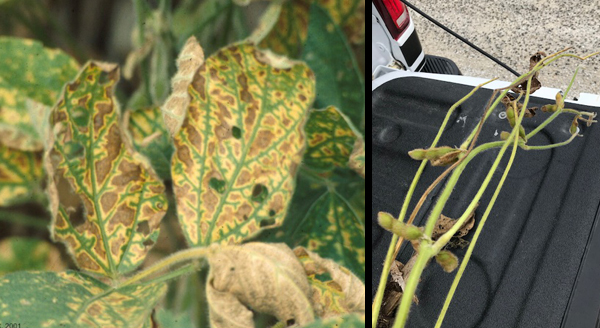
SDS. Symptoms of SDS are showing up in a few areas. Initial symptoms include yellow spots between the leaf veins that coalesce into large areas of interveinal chlorosis and necrosis (Pic. 1). However, these symptoms can also be associated with other pathogens so splitting the stems open is necessary to accurately diagnose the disease. With SDS, the internal pith will be white, while the cortex (bark) of the stem will turn milky-brown. SDS will result in affected leaves dropping while the petioles remain attached to the stem (Pic. 1) and will also cause root rot. Blue fungal structures can be found on the roots as a sign of SDS infection but they are not always found.Yield losses as a result of SDS are greater when foliar symptoms become visible during the early reproductive stages but when they show up late, yield impacts may be minimal. For more information on SDS identification and management please refer to the article here.
Brown stem rot (BSR). Another disease that can produce foliar symptoms similar to those of SDS is BSR. Splitting the stems will reveal browning of the vascular tissue and pith which does not happen with SDS. Incidence and severity of BSR is usually higher under hot dry weather during the late reproductive stages. Beware that a few soybean stems infested with Dectes Stem Borer (DSB) had an internal brown coloration where the larva had been feeding (Pic. 2) so we should not assume that an internal brown stem is BSR for sure. Check for entry points of DSB on the petiole (Pic. 2) and for any larval presence inside the stem. For more information on DSB refer to the DSB article here.

Stem canker. This pathogen can also result in foliar symptoms similar to those of SDS and BSR. However, upon closer examination, brown-reddish lesions near the nodes which expand up and down on one side of the stem (Pic. 3) can be observed. Unlike SDS, leaves of plants infected with stem canker will remain attached to the plant instead of falling off. For more information on stem canker symptoms and management refer to the article here.
Red crown rot. Foliar symptoms of red crown rot resemble those of SDS and BSR; yellow spots between the veins that grow into chlorotic areas while the veins remain green. However red crown rot can be distinguished from these other pathogens by the presence of reddish-orange structures on the stem surface near the soil line (Pic. 4). These structures are called perithecia, the reproductive structures of the fungus. Even when these structures are not visible, the lower part of the stem and roots infected with red crown rot may have a reddish discoloration (Pic. 4). This pathogen will infect the roots shortly after planting but development is favored by high moisture and high clay content.
Top dieback. An article on soybean top dieback was published here. We continue to see soybean fields with yellow and brown symptoms on the top portion of the canopy although not in an interveinal pattern like those of SDS. In one of the fields that was visited, these symptoms resemble those of potassium deficiency (Pic. 5) and in fact low K was confirmed. Those same beans also looked stunted and cysts of SCN were found on their roots. Drought conditions can lead to the development of K deficiency even when soil test shows adequate levels. When observing this type of symptoms, confirming the potential causes through testing (nutrient foliar/soil, SCN egg counts etc.) can help to more accurately diagnose the problem.


Picture 1. SDS foliar symptoms (left) and leaf drop with petiole still attached (right).
Picture 2. Dectes Stem borer larva inside soybean stem (left) and entering through petiole (right)
Picture 3. Stem canker lesions on soybean stem.
Picture 4. Red crown rot perithecia (left) and reddish coloration on soybean stem (right)
Picture 5. Soybean yellowing and top dieback symptoms.https://www.fssystem.com/Products-Services/Resource-Center/Resource-Detail/sudden-death-syndrome